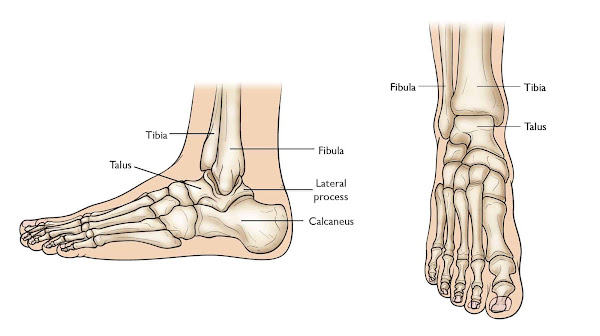
View 17 Foot And Ankle Bones Diagram - The Ankle Diagram The Ankle Joint The Bones, Tendons, Ligaments, and Muscles of the Joint The talocrual joint is composed of three major bones. The Tibia The Fibula The Talus Your body's weight is transferred from the tiba to the talus. Weight can be distributed anteriorly or posteriorly throughout the foot as a result of this. Foot and ankle bones and joints The Ankle Joint Ankle joint capsule on the lateral side. The ankle joint, also known as the tibiotalar joint, is formed when the top of the talus (the uppermost bone in the foot), the tibia (shin bone), and the fibula come together. The ankle joint is a synovial joint as well as a hinge joint. Hinge joints typically only allow for one direction of motion.
A foot pain diagram is an excellent tool for determining the source of your ankle and foot pain. There are numerous structures, such as bones, muscles, tendons, and nerves, that each produce slightly different foot pain symptoms. The Ankle and Foot The foot and ankle complex is made up of 28 bones and 25 joints. These structures are designed to accommodate the foot and ankle's stability and mobility responsibilities on various surfaces while bearing varying degrees of weight.
The Hindfoot: The hindfoot is made up of the ankle joint, which is located at the bottom of the leg and is where the tibia and fibula meet the ankle bone known as the talus. The heel bone, known as the calcaneus, is also included. The Midfoot: Our foot arches are made up of the five bones of the midfoot. They are arranged in a pyramid shape to serve as the feet's shock absorbers. With a solid understanding of foot anatomy, it is easy to determine which surgical approaches can be used to access various areas of the foot and ankle. The anatomy of the foot and ankle (Figure 1) includes a variety of anatomical structures such as bones, joints, ligaments, muscles, and tendons.
TAG : Foot And Ankle Anatomy Diagrams


